Lumbar spine

Lumbar spinal stenosis

What is it ?

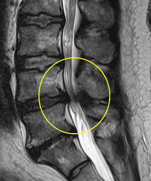
Lumbar spinal stenosis also results from disc degeneration. This causes a transfer of mechanical charges on the articular facets in particular, producing in the long term osteophytary beaks (bony outgrowths) encroaching into the lumbar canal. The volume available for the nerve roots is thus reduced. Most often it is patients over 50 who are affected. The MRI image below shows a narrowing of the L4-L5 level (yellow circle).

Clinical presentation

Decrease of the volume of the lumbar spinal canal does not necessarily cause symptoms. When symptomatic, this can manifest as low back pain and pain in the lower limbs when walking, sometimes associated with tingling (paraesthesia). All of these symptoms are called “neurogenic claudication”. The walking perimeter is limited and patients usually have fewer symptoms when they are at rest. There is clearly a dynamic effect of the symptoms.

When to consult a specialist?

When symptoms alter the quality of life, surgical treatment is discussed with the specialized spine surgeon. In fact, the enlargement of the spinal canal (decompression or laminectomy) is the most effective treatment in terms of pain relief and duration of the relief effect. Several parameters will be taken into account to decide the type of treatment, but a specialized opinion, even to discuss an injection and postpone surgery, is useful.

Non surgical treatment

Non-surgical treatment is limited when the symptoms are very disabling: painkillers, physiotherapy, infiltration, and others. However, this usually relieves for a limited time: from a few days to a few weeks most often.

Surgical treatment

The dynamic or mechanical effect of the narrow canal (stenosis), explains that it is ultimately the canal enlargement surgery that will actually be effective, as has been demonstrated by many studies comparing non-surgical treatment to surgical treatment. The main goal of surgery is enlargement of the spinal canal, but biomechanical parameters unique to each patient must be taken into account to determine whether or not instrumentation will be necessary. This in particular, is the role of sagittal balance analysis.

Postoperative follow-up

It depends on the extent of the surgery. In general, patients remain hospitalized for a few days. Rehabilitation begins the day after the operation, often at first with a walking frame, then quickly independently. For the first 6 to 8 weeks, patients experience pain from the surgery, but which is controlled by pain killers. They are encouraged to walk as much as possible and resume their daily life activities within the limits of their pain, without carrying loads. Depending on the type of surgery, it takes between 3 and 6 months to fully recover.

Risks and complications

They depend on the surgical technique chosen. There are general risks associated with any surgery. The specific risks such as serious neurological complication are very low (<1%). The infectious risk is 2 to 3%. All this is reviewed in a non-exhaustive manner with the surgeon during a pre-operative interview.